Neuroanatomy
 |
Discussion of basic and advanced neuroanatomical topics in both the central and peripheral nervous systems.
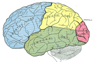
The cerebral cortex, derived from the dorsal telencephalon or pallium, consists of two hemispheres (the right and the left cerebral hemispheres), each of which is divided into five lobes. Read more »
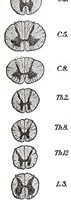
The spinal cord, along with the brain, make up the Central Nervous System (CNS). It is a thin collection of nerves that is enclosed by the vertebral column, CSF, and meninges and... Read more »
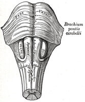
The pons, a part of the brainstem that includes the midbrain and medulla, is a bulbous structure that is 2 cm long. The pons and cerebellum together form the metencephalon. [1] Read more »
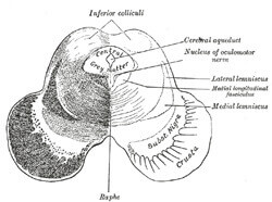
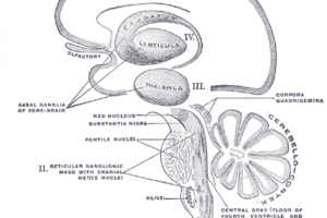
The midbrain, or mesencephalon, is positioned between the forebrain (diencephalon) and hindbrain (metencephalon)[1]. It forms the stalk-like part of the brain that connects the brain to the spinal cord. The midbrain has... Read more »
The diencephalon contains several structures, each with the term “thalamus.” Most of these structures derive from the developmental vesicle called the diencephalon. The contents of the diencephalon include the dorsal thalamus (commonly... Read more »
The thalamus is a walnut-sized structure buried at the center of the cerebrum. Nearly all sensory information that enters the cerebral cortex goes through the thalamus. The major exception to this rule... Read more »
Telencephalic areas, or cerebrum, arise from the developmental structure known as the telencephalon. These areas include the cerebral cortex, the basal ganglia, and the olfactory bulb. Together with the diencephalon, the telencephalon... Read more »
VPM and VPL send projections up to primary somatosensory cortex, defined by Brodmann’s areas 3, 1, and 2 sitting on the post-central gyrus. Because all the somatosensory tracts are crossed (remember the... Read more »
The anterolateral system carries pain and temperature information from the periphery to the VPL nucleus of the thalamus, and on up into the somatosensory cortex. Read more »
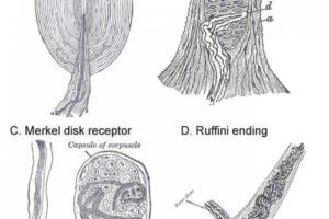
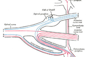
Peripheral Receptors Because this system carries a variety of sensations, including light touch, proprioception and vibratory information, it requires a variety of sensory receptor cells. The light touch and vibratory receptors are... Read more »