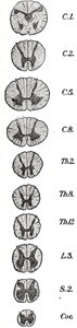
The spinal cord, along with the brain, make up the Central Nervous System (CNS). It is a thin collection of nerves that is enclosed by the vertebral column, CSF, and meninges and is a vital component in the regulation of the body. The spinal cord is located within the vertebral foramen and is made up of 31 segments: 8 cervical, 12 thoracic, 5 lumbar, and 5 sacral. A pair of spinal nerves, anterior (motor) and dorsal (sensory), exits from each segment of the spinal cord. [1]
Functions
Understanding the functions of the spinal cord are arguably the most important aspect to retain as well as correctly identifying the various conditions that may occur from spinal lesions. The spinal cord transmits somatic and autonomic sensory information from the peripheral nervous system to the brain and conducts motor information from the brain to the glands, smooth muscle, cardiac muscle, and skeletal muscles. The spinal cord controls routine behaviors like walking.
Blood pressure and temperature regulation occur through the sympathetic and parasympathetic nervous systems, the two parts of the autonomic nervous system controlled by the spinal cord. The spinal cord also acts as a minor reflex center, allowing an automatic response without engaging the brain.
The spinal nerves that are located within the spinal cord are called the Upper Motor Neurons (UMN) whereas the nerves that branch out from the spinal cord to the body are called the Lower Motor Neurons (LMN). Lesions to these neurons or spinal tracts will be discussed in other areas of the Neurobiology section.
Composition
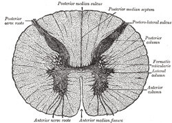
The spinal cord, like the brain, is composed of gray and white matter. The white matter consists of axons covered with a myelin sheath while the gray matter contains cell bodies and dendrites.
However, unlike the brain, the white matter is on the outer portion of the spinal cord with the gray matter in the interior.
The spinal cord is relatively short- 45 cm in men and 43 cm in women, respectively. As a result, the spinal cord does not traverse the entire vertebral column but extends as far as the last thoracic vertebrae. The spinal cord nerves that make up the rest of this distance is known as the cauda equina, or “horse tail”, due to the resemblance of its structure to a horse’s tail.
Clinical Correlations
 The spinal nerves serve to regulate the body’s functions. A lesion to any part of the spinal cord can be complete or incomplete. A complete injury means that there is no sensation or function in any area below the lesion, whereas an incomplete injury means that there may be some residual function below the lesion.
A lesion to the spinal cord at the level of the cervical spine, such as in acute C-spine trauma, may result in complete paralysis of all four limbs (quadriplegia). A lesion above the C4 spinal nerve may result in inability to inspire. The immediate goal of management in spinal cord injury is preventing neurological injury due to further movement as well as treating any cardiovascular or respiratory instability.
The spinal cord may be injured by compression from bone, ligaments, edema, overstretching of neural tissue, circulation disruption, and direct injury. [2]
Other causes of spinal cord conditions:
- Tumors
- Infections such as meningitis and poliomyelitis
- Inflammatory diseases
- Autoimmune diseases
- Degenerative diseases such as amyotrophic lateral sclerosis and spinal muscular atrophy
References
Nervous System – Spinal Cord. BBC News. http://www.bbc.co.uk/science/humanbody/body/factfiles/spinalcord/spinal_cord.shtml
National Institute of Neurological Disorders and Stroke. http://www.ninds.nih.gov/disorders/brainandspinaltumors/brainandspinaltumors.htm