Peripheral Receptors
Because this system carries a variety of sensations, including light touch, proprioception and vibratory information, it requires a variety of sensory receptor cells. The light touch and vibratory receptors are in the skin, and there are four types in glaborous skin (skin without hair).
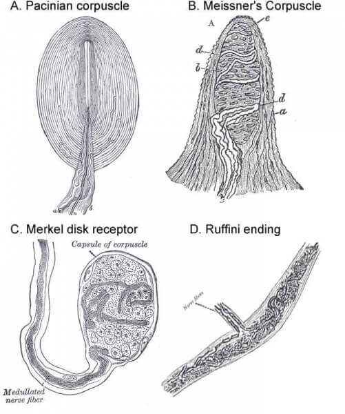
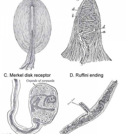
Pacinian Corpuscles
Pacinian corpuscles sit deep in the dermis and sense vibratory stimuli. They typically respond to the beginning and end of a constant stimulus, but very quickly adapt to constant stimuli. Because the respond to changes in pressure, rather than constant pressure, they respond well to vibrations. However, because they are found deep in the dermis Pacinian corpuscles do not have good spatial resolution.
Meissner’s Corpuscles
Like the Pacinian corpuscles, the nerve endings of Meissner’s corpuscles are embedded in an extracellular matrix that makes them more sensitive to vibratory stimuli than constant stimuli. However, because they are more superficial than Pacinian corpuscles, sitting near the epidermis-dermis border, they have better spatial resolution.
Merkel Disk Receptors
Merkel disks consist of sensory receptor cells coupled, probably by a chemical synapse, to a sensory neuron. While the exact mechanism of mechano-sensory transduction is unknown, we can guess that deformation of the skin exerts pressure on the epithelial cell of the Merkel disk through the tight junctions it shares with neighboring epithelial cells. The cell surrounds its associated nerve ending and probably releases some sort of neurotransmitter onto the nerve, which can fire an action potential up.
Because the Merkel disk receptors sit superficially and are tightly coupled to basal epithelial cells, they are in a position to sense light touch with excellent spatial resolution. The density of this type of receptor is particularly high in parts of the body, like the fingertips, for which fine touch sensation is important.
The Merkel disk receptors adapt slowly, so they continue to signal constant stimuli much longer than either Pacinian or Meissner’s corpuscles.
Ruffini Endings
Like Merkel disks, Ruffini endings signal constant stimuli well. However, because they are found deeper in dermis than Merkel disks, Ruffini endings have poor spatial resolution. Consistent with that, the density of Ruffini endings is almost always less than that of Merkel cells.
Receptor Density Varies by Area
Our fingertips are much more sensitive to touch then is our back, and this is due primarily to differences in receptor density between the two areas. The density of all four types of receptors is much higher in the fingertips than in the back. The easiest way to appreciate this difference is to try the two-point discrimination test, for which you can use the backs of a pair of pins to test how far apart the pins need to be to tell if there are two or only one pin on different parts of the skin. At the fingertips, you can sense two separate points at distances as small as 2 mm. Up on the arm, you might not be able to distinguish two pins even if they are separated by 20 mm, and on the back the minimum distance can be greater.
Peripheral Nerves Innervate Dermatomes

Because peripheral nerves enter the spinal cord through the dorsal root entry zone at each spinal level, each peripheral nerve carries information from a restricted band of skin. This band is called a dermatome, and mapping out sensory deficits by dermatome can help localize lesions.
Cell Bodies of Peripheral Nerves in Dorsal Root Ganglion
All the somatosensory neurons from the body have their cell bodies in the dorsal root ganglion, and extend an axon into the spinal cord through the dorsal root entry zone.
Dorsal Column Tracts
Axons from the somatosensory neurons of the DC-ML system travel rostrally in the dorsal funiculus, which contains two major tracts: the fasciculus gracilis and fasciculus cuneatus.
 Fascicullus Gracilis
The fasciculus gracilis carries fibers from the lower body, including the legs. As fibers enter, they are added to the lateral side of the dorsal funiculus, so the most medial fibers in the fasciculus gracilis come from the lowest dermatome. Because fibers are coursing rostrally, all levels of the spinal cord have a fasciculus gracilis.
Fascicullus Cuneatus
The fasciculus cuneatus carries fibers from the upper half of the body, including the arms. It is the more lateral of the two fasciculi in the dorsal funiculus, and, because it carries fibers from the arm, is only found at cervical levels.
Nuclei and Decussation in the Medulla
Fibers from both fasciculus gracilis and cuneatus terminate and synapse within the medulla in nucleus gracilis and nucleus cuneatus, respectively. The second order neurons, whose dendrites sit in the nuclei, the decussate immediately through the internal arcuate fibers. Recall that when fibers decussate they typically cross both the midline (hence “decussation”) and the dorsal-ventral boundary. So while the primary sensory neurons run in the dorsal half of the spinal cord (like good sensory systems should), the second-order decussated fibers move into the ventral portion of the medulla to run in the medial lemniscus.
Medial Lemniscus Carries Fibers to Thalamus
Medial lemniscus starts medial in medulla, then as it courses rostrally its position shifts laterally around the bottom of the tegmentum. Its final position is lateral as it enters the VPL of the thalamus. As it moves rostrally the medial lemniscus picks up fibers from the trigeminal nuclei, with the fibers adding to the medial/ventral end of the medial lemniscus.
The medial lemniscus is somatotopically organized, meaning that its organization can be mapped onto the shape of the body. Nearby parts of the body are represented by nearby locations in the medial lemniscus.
VPL is the Primary Thalamic Relay Nucleus for Somatosensation from the Body
The medial lemniscus terminates in the ventral-posterior-lateral nucleus of the dorsal thalamus. The is the primary relay nucleus of the somatosensory system for the body, receiving input from both the dorsal column-medial lemniscus system and the antero-lateral system. The VPL then projects out to the primary somatosensory cortex through the internal capsule.