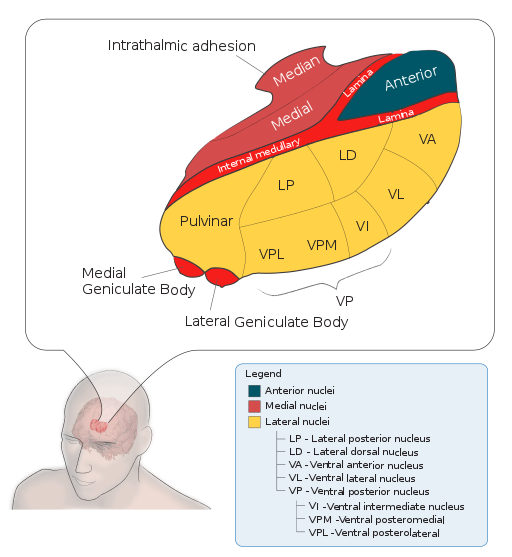
The thalamus is a walnut-sized structure buried at the center of the cerebrum. Nearly all sensory information that enters the cerebral cortex goes through the thalamus. The major exception to this rule is olfactory information, which goes from the olfactory epithelium to the olfactory bulb and directly to cortex (piriform cortex is a main olfactory target).
The role of the thalamus has been unclear. It relays information from the periphery to cortex, but it does not seem to modify the information. Since there is no apparent reason to have a relay nucleus, the thalamus had been ignored for decades. In fact, the thalamus seems to play a major role in regulating information flow – suppressing sensory input to cortex during sleep, and allowing input during wakefulness.
Connections
Sensory input comes to thalamic relay nuclei through a variety of pathways. For example, visual inputs to the LGN (lateral geniculate nucleus) arrive via the optic tract. Inputs to the MGN arrive through the brachium of the inferior colliculus. Inputs to VPM (ventral posterior medial nucleus) and VPL (ventral posterior lateral nucleus) arrive from the medial lemniscus. The output of relay nuclei project to cerebral cortex. These glutamatergic (excitatory) outputs project through the ventral thalamus (TRN – thalamic reticular nucleus), and give off collaterals that synapse within the TRN.
The bulk of the axons reach cortex through the internal capsule. The internal capsule flows into the corona radiata, the posterior portion of which projects to the visual cortex and is called the optic radiations
Connections from cortex back to the thalamus also travel through the internal capsule. Connections from cortical layer VI terminate in primary relay nuclei and appear to modulate what information gets relayed to cortex. Connections from cortical layer V project to higher order thalamic nuclei, like the pulvinar which is the higher order nucleus for the visual system.
Blood Supply to the Thalamus
Most of the thalamus is supplied by branches of the posterior cerebral artery.
The LGN is supplied by at least 2 arteries: “The lateral geniculate body has a dual blood supply from the anterior choroidal artery (branch from internal carotid artery) and from the lateral choroidal artery (branch from the posterior cerebral artery).” (Journal of Neurology, Neurosurgery and Psychiatry)
Structures to Identify on Sections
To have a good handle on the thalamus, you should be able to identify all the structures shown in Nolte pp. 393-396. The highest yield nuclei include:
- LGN – dorsal lateral geniculate nucleus is the primary visual relay nucleus. Mnemonic: Light geniculate nucleus.
- MGN – medial geniculate nucleus is the primary auditory relay nucleus. Mnemonic: Musical geniculate nucleus.
- VPM – ventral posterior medial nucleus, the primary relay nucleus for somatosensory information from the face. Mnemonic: VP Mirror nucleus to see your face.
- VPL – ventral posterior lateral nucleus, the primary relay nucleus for somatosensory information from the body.
References
The most authoritative online reference of thalamus is written by S. Murray Sherman, of Neurobiology at the University of Chicago.
J. Nolte (2002) The Human Brain: An introduction to its functional anatomy. 5th edition. Mosby, Inc. St. Louis. Ch. 16.
C Luco, A Hoppe, M Schweitzer, X Vicuna and A Fantin (1992) Visual field defects in vascular lesions of the lateral geniculate body. Journal of Neurology, Neurosurgery, and Psychiatry. 55:12-15.


