The pons, a part of the brainstem that includes the midbrain and medulla, is a bulbous structure that is 2 cm long. The pons and cerebellum together form the metencephalon. [1]
Organization
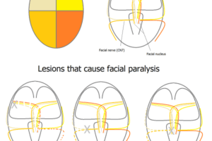
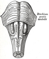
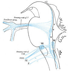
The pons is below the midbrain and in front of the cerebellum. It is formed from nerves that travel transversely, rather than vertically as is the case with the majority of brainstem structures. Many cranial nerves exit the pons or have their nuclei in the pons. These include cranial nerves V, VI, VII, and VIII. The fourth ventricle of the brain is formed from the posterior pontine surface.
The pontine arteries, branches of the basilar artery, provide the majority of the blood supply to the pons. The anterior inferior artery and superior cerebellar artery also play a role in perfusion.
The pons contains motor and sensory neuronal pathways that come from the cerebrum, spinal cord, and cerebellum.
Function
 The pons is an integral part of the brain’s pathways. It is an important sensory relay system that provides information to the cerebellum, cerebrum, and spinal cord. [2] The pons provides input to the cerebellar cortex through the pontine nuclei, allowing the cerebellum to coordinate much of its control.
The pons also functions as a motor relay center. Many of the descending nerve fibers synapse in the pons. As a result, any injury to the pons may result in motor deficits.
The pons is also an important control center for respiration. The apneustic center, located in the lower pons, stimulates inspiration and the pneumotaxic center, located in the upper pons, inhibits inspiration by decreasing the activity of the phrenic nerve. If the pneumotaxic center were damaged, inspiration would be prolonged and there would be a decrease in the respiratory rate.
Some experts believe that the pons may play a role in REM sleep and in arousal.
Due to the fact that the pons contains several cranial nuclei or nerve origins, the pons also functions to relay information from the face, teeth, ear, and eyes and their subsequent adjustment.
Clinical Correlation
 Conditions that affect the pons may be detrimental. The most famous condition is known as central pontine myelinosis[3] which is caused by damage to the myelin sheaths of the brainstem nerve cells. It may present as difficulty in speaking but may also present as a “locked-in syndrome” in which the patient is completely aware of what is going on in the surrounding environment but the body is completely paralyzed except for the eye muscles.
Central pontine myelinosis is often caused by rapid correction of hyponatremia, which is a low level of sodium in the blood [the normal range for sodium is 135-145 milliequivalents per liter]. Slow correction of a hyponatremic condition is necessary to prevent this from occurring.
Central pontine myelionosis is diagnosed by MRI.
Tumors may also occur in the pons. These are usually found in children around the age of six years old.
Cranial nerve VI, or abducens, is the most commonly damaged cranial nerve. Damage to this nerve results in ipsilateral gaze palsy.
References
Pons. Wikipedia. http://en.wikipedia.org/wiki/Pons
Brainstem- Pons. Union County College. http://faculty.ucc.edu/biology-potter/the_brain/sld017.htm
Vellaichamy, Muthukumar. Hyponatremia. Emedicine. http://www.emedicine.com/PED/topic1124.htm