The midbrain, or mesencephalon, is positioned between the forebrain (diencephalon) and hindbrain (metencephalon)[1]. It forms the stalk-like part of the brain that connects the brain to the spinal cord. The midbrain has been described as archipallian in origin, meaning that it is mediated by aggression or submission- both very primitive instincts.
Organization
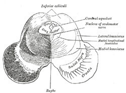
The mesencephalon is essentially divided into the tectum (meaning “roof”) and cerebral peduncle. The tectum is further divided into inferior colliculi and superior colliculi. The superior colliculi is involved in visual functions while the inferior colliculi is involved in auditory functions. The fourth cranial nerve, known as the trochlear nerve, exits the brain below the inferior colliculus.
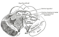 The cerebral peduncle (or crus cerebri in Latin), which forms the anterior half of the midbrain, contains the midbrain tegmentum, crus cerebri, and substantia nigra (this produces the neurotransmitter dopamine and has been implicated in Parkinson’s disease). The cerebral peduncle is a band of neurons that the brain’s motor information to the body- the corticospinal and corticobulbar tracts run through the cerebral peduncles. For the most part, the cerebral peduncles are covered by the temporal lobes of the cerebrum. The third cranial nerve, the oculomotor nerve, exits the brain between the peduncles.
The midbrain contains the cerebral aqueduct, which communicates between the third and fourth ventricle.
The substantia nigra contains multipolar nerve cells and is usually identified by its dark pigmentation. it is divided into the substantia nigra pars compacta and the substantia nigra pars reticulata. Substantia nigra pars compacta is the one that is necessary for dopamine production and transmits information to and from the caudate and putamen.
The red nucleus is part of the rostral midbrain and is involved in the control of movement through the rubrospinal tract. [2] The red nucleus is theorized to have a high iron content and to have especially high vascularity.
Functions
The midbrain controls many important functions in the brain. It is responsible for eye movement, visual and auditory systems, body movement, and pupil dilation. It does so by routing information from the environment to the cerebral cortex. The midbrain also plays an endocrine role in arousal of the autonomic system and cortex.
The rubrospinal tract forms a path between the cerebellum to the lower motor centers and is important as an another route for mediation of voluntary movement, especially in causing flexion and inhibiting extension of the upper extremities. In the spinal cord, it accompanies the lateral corticospinal tract and has been known to replace many of the duties of the corticospinal tract in some primates when there is damage to the corticospinal tract.
The substantia nigra is important in regulating mood and controlling voluntary movement.
Clinical Correlation — Parkinson’s Disease
Parkinson’s disease is a degenerative CNS disorder that causes muscle stiffness, unintentional tremor, poor balance, and slowing of movement. It also falls under the classification of movement disorder. It is caused by lack of stimulation of the motor cortex due to the degeneration of the substantia nigra which produces dopamine[3].
References
Mesencephalon. Wikipedia. http://en.wikipedia.org/wiki/Midbrain
Midbrain. Brain Atlas. Lundbeck Institute. http://www.brainexplorer.org/brain_atlas/Brainatlas_Midbrain.shtml