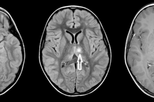
Tumefactive multiple sclerosis (MS) is an unusual but important manifestation of multiple sclerosis.
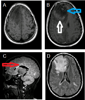
This is a patient with tumefactive MS. Notice the incomplete rim enhancement around the right frontal mass on the T1 post contrast sequence. This incomplete rim enhancement is classic for a tumefactive demyelinating lesion as opposed to a neoplasm. A. T1 precontrast and B. postcontrast images demonstrate a T1 hypointense right frontal mass with incomplete rim enhancement (white arrow) and right to left subfalcine herniation (blue arrow). C. Sagittal and D. axial FLAIR images demonstrate hyperintense lesions involving the pericallosum (red arrow) and left temporal subcortical white matter.
With an incidence of 1-2 per 1000 cases diagnosed, tumefactive MS is generally defined as a large demyelinating lesion that mimicks a neoplasm. Typically these lesions are solitary and greater than 2 cm. Clinical presentation is non-specific and includes a spectrum of symptoms ranging from confusion to focal motor signs depending on the location of the lesion.
MS is the “D” in MAGIC Dr a helpful mnemonic to remember the primary causes of a rim enhancing brain mass: Metastasis, Abscess, Glioblastoma Multiforme, Infarct, Contusion (trauma), Demyelinating.
Given their overlapping features it is often hard to distinguish tumefactive MS from tumor although specific imaging findings favor MS. One important finding is the presence of incomplete rim enhancement. Tumors typically manifest complete rim enhancement. Other less specific findings favoring tumefactive MS include T2 isointensity in the region of enhancement, size out of proportion to vasogenic edema and secondary signs of MS such as pericallosal lesions
Establishing a diagnosis is important as the treatment for tumefactive MS differs from other etiologies of rim enhancing mass lesions in the brain. LP may be contraindicated if the lesion is large. Biopsy is the gold standard for diagnosis, and will usually differentiate this demyelinating lesion from a neuro-oncology disorder.
Selected References
Kaeser MA, Scali F, Lanzisera FP, Bub GA, Kettner NW. Tumefactive multiple sclerosis: an uncommon diagnostic challenge. J Chiropr Med. 2011 Mar;10(1):29-35.