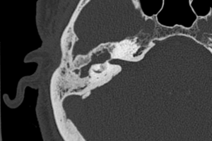
There are three semicircular canals – superior, lateral and posterior. The semicircular canals are integral to maintaining balance and equilibrium with changes in body and head position. Dehiscence of the superior semicircular canal is a rare finding characterized by a partial absence of the superior aspect of the otic capsule, resulting in a communication between the semicircular canal and the intracranial contents.
Diagnosis: Dehiscence of the right superior semicircular canal
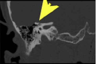
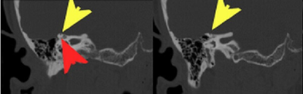
Figure 1: Coronal reformats from a temporal bone CT (using a bone reconstruction algorithm with thin sections through the right internal auditory canal) demonstrate absence of bone (yellow arrow) covering the roof of the superior surface of the semicircular canal (red arrow). This finding is pathogmnemonic for dehiscence of the superior semicircular canal.
This is best appreciated on the reformatted images from an internal auditory canal protocol CT – coronal and semioblique, sagittal and coronal reformats will show the absence of the superior aspect of the otic capsule on more than one slice. The diagnosis can be challenging however because it is excluded by even a thin bony roof that can exist at or below the resolution of modern CT or be masked by partial volume averaging with adjacent slices
Classic presentation of the syndrome of superior semicircular canal dehiscence is Tulio’s phenomena – sudden sounds induce vertigo and/or nystagmus. Other symptoms include autophony (reverberation when speaking), sensorineural hearing loss and vertigo exacerbated by valsalva.
Treatment of superior semicircular canal dehiscence involves craniotomy with middle cranial fossa repair through bony resurfacing.
Selected References
Swartz J, Loevner L. Imaging of the Temporal Bone. 4th Ed. Thieme New York 2009