Masses of the pineal region can be divided into pineal and non pineal. Non pineal masses can arise from any of the structures in the region and include glioma (from the tectum or corpus callosum), meningioma (from the dura), epidermoid and arachnoid cysts (from the CSF) and lipoma (from the interhemispheric fissure). Other processes, like cerebral edema or intracranial hypotension can efface the pineal region, but they are not specific to this area.
Diagnosis: Tectal plate glioma
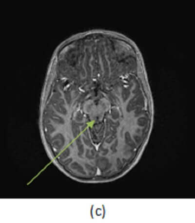
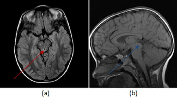
Figure 1: (a,red arrow) FLAIR hyperintense signal expands the right tectal plate with T1 isointense fullness in the same location (b,blue arrow) that does not enchance (c, green arrow).
Tectal plate gliomas are aggressive, infiltrating malignancies of the tectum (the dorsal midbrain). They are generally expansile T2/FLAIR hyperintense minimally enhancing lesions that expand the tectum and compress the midbrain leading to hydrocephalus. They most commonly present as WHO grade 3 (anaplastic astrocytoma) tumors, but may undergo secondary transformation into a glioblastoma multiforme (GBM). Treatment involves resection with adjuvant chemo and radiation therapy but prognosis is poor, with a median survival of 2-3 years.