Imaging plays an important role in the diagnosis and management of patients with primary brain neoplasms. In particular, imaging is typically performed within 48 hours of tumor resection.
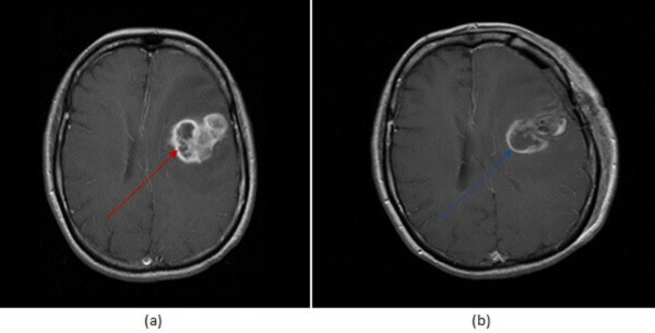
Diagnosis: Residual GBM after surgery
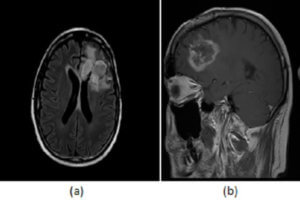
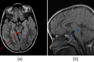
Figure 1: (a, red arrrow) Pre and immediate post operative (b, blue arrow) MRI imaging of a GBM demonstrates decreased but persistent enhancement within the surgical bed, consistent with recurrent tumor . Granulation tissue takes more than 48 hours to develop.
The goals of imaging at this timepoint are to:
1. Assess for residual tumor and establish a new imaging baseline.
- Since granulation tissue takes more than 48 hours to form, mass like enhancement in the surgical bed in immediate post operative imaging is consistent with residual tumor.
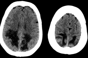
2. Look for signs of infarct.
- While non territorial infarcts are common after surgery, they are important to identify because subacute infarcts can enhance for months and therefore mimic tumor recurrence on subsequent imaging.
3. Recognize expected post operative changes:
- DWI : Ischemia/infarct due to retraction, small vessel damage,
- Pneumocephalus, and
- Small amount of intra and/or extra axial blood (mimic of enhancement).
4. Look for unexpected post operative complications:
- Territorial ischemia,
- Large intracerebral hemorrhage,
- Venous sinus thrombosis,
- Hydrocephalus, and
- CSF leak pseudomeningocele.