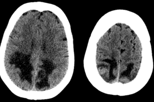
The differential diagnosis for an intra-axial posterior fossa mass depends on the age of the patient. The most common causes are juvenile pilocytic astrocytoma, medulloblastoma and ependymoma in a child and metastasis and hemangioblastoma in an adult, with the former being much more common. Be sure to exclude complications of posterior fossa masses in all patients, including obstruction of the fourth ventricle leading to hydrocephalus and herniation of the cerebellar tonsils below the foramen magnum.
Diagnosis: Metastatic lung cancer to the cerebellum

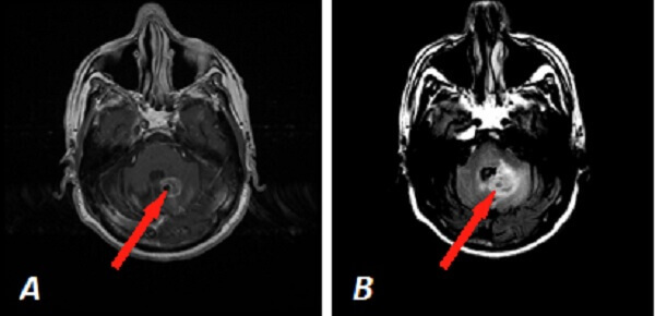
Figure 1: Red arrow (a) and (b) Axial T1 post contrast and FLAIR images demonstrate a circumscribed intra axial enhancing mass in the left cerebellar hemisphere with surrounding vasogenic edema abutting the fourth ventricle. (C) 3D rotational MIP from a PET-CT scan demonstrates hypermetabolic right upper lobe pulmonary nodules consistent with lung cancer (in this case non small cell).
Brain metastasis are the most common intraaxial neoplasm in adults, occuring in 8-10% of cancer patients. Lung, breast and melanoma are the most common primaries. Brain metastasis are especially common in small cell lung cancer (80% of patients 2 years after diagnosis, compared with 30% in non small cell lung cancer)1. Treatment commonly includes steroids (to reduce mass effect) and whole brain radiation.
References
1Chi A, Komaki R. Treatment of Brain Metastasis from Lung Cancer. Cancers 2010, 2, 2100-2137