Acute infarcts lead to cell swelling in a process called cytotoxic edema. Signal in diffusion weighted MRI (DWI) is inversely related to the degree of brownian motion of intraparenchymal water. When an infarct produces cytotoxic edema, water is trapped inside the cell results in increased DWI signal. Sometimes, increased DWI can be an artifact of high T2/FLAIR signal, an artifact also known as shine through – to eliminate the T2 “shine through” effect on DWI, an apparent restricted diffusion (ADC) map is created.
Low signal on ADC represents real restricted diffusion. While real restricted diffusion is most commonly seen with acute infarcts (as in this case), it can be seen with a variety of other conditions such as seizures, abscesses, highly cellular tumors, hemorrhage and hypoglycemia.
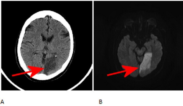
Diagnosis: Acute left posterior cerebral artery stroke.

Figure 1: Red arrows. A. Axial non-contrast head CT demonstrates hypoattenuation of the left medial occipital lobe with loss of grey white differentiation. B and C. Axial diffusion weighted (DWI) and apparent diffusion coefficient (ADC) images demonstrate respectively increased and decreased signal in the occipital cortex.
The paired posterior cerebral arteries supply portions fo the thalamus, lateral ventricle, midbrain occipital and inferomedial temporal lobes. Patients with acute PCA territory infarcts typically experience visual symptoms which can range from honomymous hemianopsia to cortical blindness depending on the extent of the stroke.