Parotid masses are often seen incidentally on CT scans of the brain and neck. The most common primary parotid mass is the benign mixed tumor (also known as a pleomorphic adenoma). These masses are hyperintense on T2 and well circumscribed.
Diagnosis: Parotid lymphoma
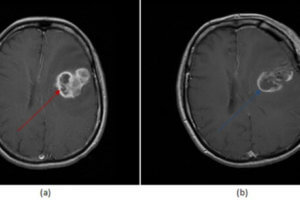
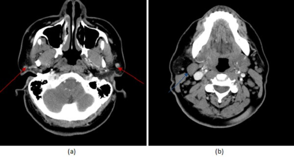
Figure 1: (a, red arrow) axial neck CT with contrast demonstrates small homogenously enhancing well circumscribed left parotid masses associated with an enlarged level 2 lymph node (b, blue arrow).
It is difficult to distinguish benign from malignant parotid masses based on imaging alone. Benign appearing lesions (such as in this case) can be malignant. Features suggestive of malignancy include ill defined borders, facial nerve paralysis, T2 signal and multifocality and lymphadenopathy. Ultrasound guided biopsy is often required to establish the diagnosis of a parotid mass.