Paragangliomas are rare neuroendocrine tumors that originate In the glomus cells of the sympathetic nervous system. The most common location is the carotid space, either involving the carotid body (glomus cells involved in homeostasis sensitive to the partial pressure of oxygen in the blood) or the vagus nerve.
Diagnosis: Paraganglioma (glomus vagale)
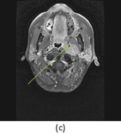
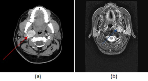
Figure 1: (a,red arrow) contrast enhanced neck CT demonstrates a homogenously enhancing carotid space mass that displaces the right ICA anteromedially. MR soft tissue neck from a different patient with the same diagnosis show a T2 hyperintense (b, blue arrow) mass that homogenously enhances with the exception of a few low attenuation foci within the mass (c,green arrow) suggestive of small flow voids. These findings are consistent with a paraganglioma of the glomus vagale.
These locations can be distinguished by the displacement of adjacent structures. The carotid body paraganglioma will splay the ECA and ICA, while the glomus vagale will displace the ICA antero-medially and the parapharyngeal fat anteriorly. Paragangliomas can also be found next to the cochlear promontory of the middle ear (glomus tympanicum) and in the jugular fossa (glomus jugulare).
Paraganglioms are avidly enhancing tumors that have a characteristic salt and pepper appearance on MRI due to the presence of vascular flow voids interspersed within enhancing tumor parenchyma.
Most glomus paragangliomas are asymptomatic or present with vocal cord paralysis due to vagal nerve involvement. Depending on their size or symptoms, these tumors are either resected (often after embolization given their vascular nature) or observed.