The differential diagnosis for an orbital mass is extensive and includes both benign (cavernous hemangioma, sarcoid, varix, meningioma and pseudotumor) and malignant (orbital lymphoma, metastasis, glioma) lesions.
Diagnosis: Orbital lymphoma
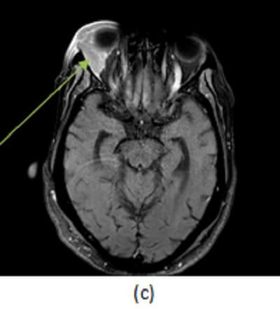
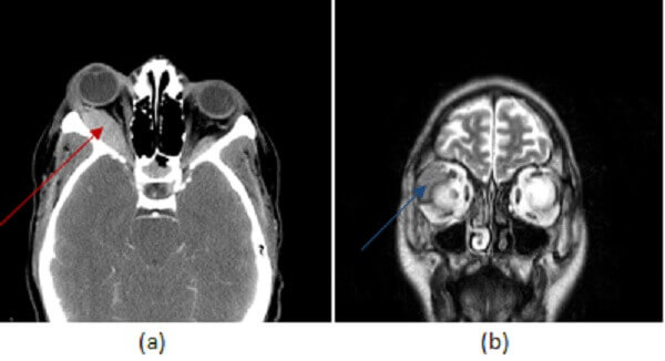
Figure 1: (a,red arrow) post contrast head CT demonstrates a smoothly marginated homogenously hyperdense extraconal mass, inseparable from the adjacent lateral rectus muscle. (b,blue arrow) Coronal T2 and (c, green arrow) axial postcontrast fat saturated T1 brain MR images in another patient with lymphoma demonstrate a tubular homogenously T2 hypointense and enhancing mass in the right lateral extraconal space that incorporates the lacrimal gland. These findings are most consistent with orbital lymphoma given the history.
Cavernous hemangioma is the most common orbital lesion. It is painless well circumscribed, homogenously hyperdense on CT, heterogeneously enhancing and T2 hyperintense with chemical shift artifact (due to fat content within the hemangioma) on MRI, orbital pseudotumor is the most common painful orbital mass, has a variety of appearances and can involve almost any orbital structure. It is characterized by inflammatory stranding of the orbital fat, heterogenous enhancement and decreased T2 signal relative to most other orbital lesions.
The imaging characteristics of orbital pseudotumor are similar to orbital lymphoma – history (of lymphoma), onset (less acute) and symptoms (less painful) can be helpful clinical findings to favor one diagnosis over the other, although biopsy is required for definitive diagnosis. Sometimes an orbital lesion can be the presenting finding of systemic lymphoma and location predicts this risk – patients with bilateral orbital lesions, and those with lesions involving preseptal structures including eyelid and orbit are more predisposed to systematic disease.
Treatment depends on the grade of the lymphoma present. Low grade disease often responds to radiation whereas high grade requires chemotherapy or immunotherapy. When orbital lymphoma is suspected, it is important to do whole body imaging such as with PET-CT to evaluate for systemic disease.