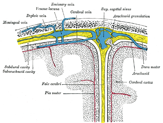
The meninges are made up of three layers (dura mater, arachnoid, and pia mater) that enclose the central nervous system. Working with the cerebrospinal fluid, they function to cushion and nourish the brain and spinal cord. The brain and cerebrospinal fluid both have the same specific gravity, so any trauma to the head causes uniform displacement of the brain, distributing any ill effects.
The word mater means “mother” in Latin and refers to the shielding nature of the meninges.
Dura Mater
The dura mater (“hard mother” in Latin) is a very tough and inflexible membrane that is composed of a superficial periosteal layer and a deep meningeal layer. The superficial layer serves as the internal periosteum of the cranium. It often travels with the deep meningeal layer of the dura but is sometimes separated by sinuses called dural venous sinuses. These sinuses function to drain blood and cerebrospinal fluid away from the brain and then through the internal jugular vein -> subclavian vein -> brachiocephalic vein -> superior vena cava -> right atrium of the heart.
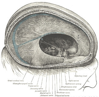 The dura mater’s meningeal layer also form septa which divide the brain into two lateral compartments and also creates a posterior compartment for the cerebellum. These septa are known as the falx cerebri and the tentorium cerebelli, respectively.
The spinal dura mater is not as tightly attached to the spinal cord as it is to the cranium and exists only as the meningeal layer. It attaches at the level of the 2nd sacral vertebrae.
The dura mater is a mesoderm derivative.
Arachnoid Mater
The arachnoid mater (cob web like in Latin) is the thin, transparent, delicate, avascular, mesh-like membrane that exists between the dura and pia maters and covers both the brain as well as the spinal cord. Like the dura, it plays a major role in cushioning of the brain and prevention of damage due to trauma. A potential subdural space exists between the dura and the arachnoid, however in living patients the arachnoid layer pushes through the dura and forms arachnoid granulations. Arachnoid granulations carry cerebrospinal fluid to the sinuses where they will be drained.[1]
The arachnoid and pia maters are often grouped together under the heading of leptomeninges. The area in between these two layers is referred to as the subarachnoid space- the cerebrospinal fluid flows here.[2]
Pia Mater
The pia mater (meaning tender mother in Latin) is the innermost layer of the meninges. It is a thin, vascular, transparent, collagenous membrane that is highly adherent- it follows the contours of the brain, including its gyri and sulci. The pia forms a constituent of the blood-brain barrier. Along with the ependyma, it forms the choroid plexus which makes the cerebrospinal fluid. Beneath the pia mater is the perivascular space which functions as a lymphatic system of the brain. The brain has no true lymphatic drainage.
The pia mater, along with the arachnoid, is a neural crest derivative.
Clinical
Meningitis
Meningitis is an inflammation of the meninges of the brain and spinal cord. It is usually caused by bacteria and viruses but may be caused by fungi. The most common bacterial causes of meningitis are Streptococcus pneumoniae or Neisseria meningitidis. The most common viral cause of meningitis are the Enteroviruses.
Symptoms of meningitis may include
- Nuchal rigidity, which is neck stiffness that prevents flexion
- Hyperreflexia
- Fever
- Severe headaches
- Nausea
- Coma
The leptomeninges are most often affected by meningitis.
Hemorrhage
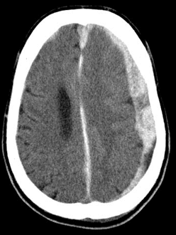
Epidural hematoma is often due to trauma that causes an accumulation of blood between the dura mater and the skull when the middle meningeal artery is injured. On CT scan, the appearance of the hematoma has been described as “lentiform”. Also, there is often a lucid interval after trauma in which the patient regains consciousness temporarily.
Subarachnoid hemorrhage is often due to rupture of a brain aneurysm. It is typically described as the patient’s “worst headache” and its onset is likened to a thunderclap.

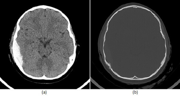
Subdural hematoma is common in chronic alcoholics as well as the elderly after a fall. It is often due to laceration of the bridging veins that connect the dura and arachnoid. The CT scan appearance of the hematoma is “crescent-shaped”.
Other meningeal problems that may arise include meningiomas, which are tumors that develop from the meninges.
References
[1]CSU, Chico, Patrick McCaffrey, Ph.D. Neuroanatomy of Speech, Swallowing and Language. http://www.csuchico.edu/~pmccaffrey//syllabi/CMSD%20320/362unit3.html
[2]Paulo Luzio Marques Araujo. Neuroanatomy. http://www.medstudents.com.br/basic/neuran/neuran2.htm