Situated in the midline just above the skull base and bridging the sphenoid and temporal bones, the cavernous sinus is a collection of venous channels that serves as a major conduit for venous drainage in the brain. For instance, the cavernous sinus receives venous drainage from superficial cortical veins and the superior ophthalmic vein, draining into the basilar plexus posteriorly along the clivus. The cavernous sinus is also connected to the transverse and sigmoid sinuses through the superior and inferior petrosal vein.
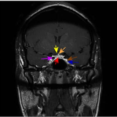
Figure 1: Some basic cavernous sinus region anatomy, with arrow color designated in brackets. Cavernous sinus (blue), optic chiasm (yellow), pituitary gland (red), infundibulum (orange), cavernous carotid (purple), intra cavernous portion of cranial nerves 3-5 (brown).
The cavernous sinus also contains several critical structures – the internal carotid arteries, cranial nerves 3, 4, 6 and the first two branches of 5. Most of the cranial nerves run along the wall of the sinus which is formed by dural reflections at the skull base. Only cranial nerve 6 runs within the cavernous sinus and as such is the most likely nerve to be affected by cavernous sinus pathology.
As the cavernous sinus is partly responsible for venous facial drainage, facial infection can result in cavernous sinus thrombosis, a potentially devastating condition. The normal cavernous sinus should enhance avidly on MRI – any lack of enhancement should raise the question of thrombosis.