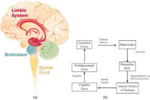
A number of pathological processes affect the limbic system. Patients with longstanding temporal lobe seizures can develop mesial temporal sclerosis, a condition characterized by atrophy of the hippocampus. Neoplasms, both benign and malignant, can also affect the limbic system . Alzheimer’s causes the deposition of beta amyloid beta plaque within the neurons of multiple limbic structures, like the entorhinal cortex and the hippocampus, and herpes simplex encephalitis has a predilection for the limbic system. .
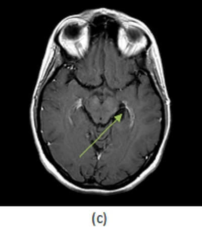
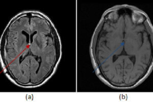
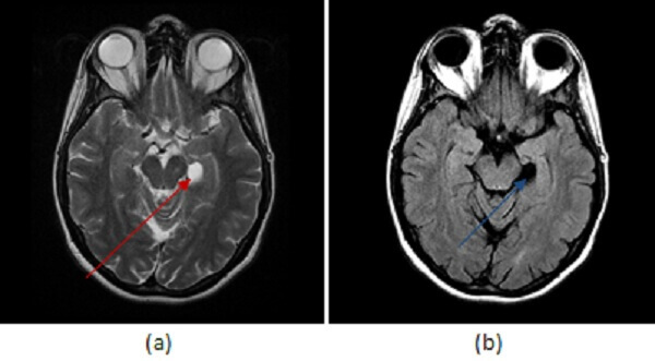
Figure 1: (a) axial T2 demonstrates a circumscribed T2 hyperintense lesion (red arrow) without increased FLAIR signal (b, blue arrow) or contrast enhancement (c, green arrow).
Some benign congenital variants of the limbic system can mimic real pathology and it is important to be aware of such variations to avoid mistaking them for disease. For example, choroidal fissure cysts are benign extraaxial cysts (typically arachnoid or neuroepithelial) that arise within the choroidal fissure, a c shaped cleft that separates the medial border of the temporal lobe from the diencephalon. Choroidal fissue cysts are incidental asymptomatic findings that require no further imaging or evaluation. The differential diagnosis includes low grade cystic neoplasms such as dysembryoplastic neuroepithelial tumor (DNET) or a ganglioglioma, but former typically has a soap bubble appearance, the later may have a mural nodule or enhance and both should have some degree FLAIR signal abnormality.
A simple cystic lesion in the hippocampal sulcus without enhancement or FLAIR signal abnormality is a choroidal fissure cyst and should not be mistaken for limbic system pathology.