Functional MRI (fMRI) is a new imaging modality that allows for dynamic evaluation of brain function in addition to structure. In the brain, blood flow is tightly coupled to neuron function.
Neurons depend on local blood supply for glucose, so when neurons fire, the local blood flow is increased to provide the glucose necessary for their activation. While MRI cannot detect the activation of neurons per se, it is sensitive to changes in blood flow. Functional MRI uses these subtle changes in blood flow as a proxy for neuron function.
FMRI has found clinical use in pre-surgical planning for brain tumor resection. Resection of tumors that abut eloquent cortex (those cortical areas vital for sensory, motor and language function) carry a high risk of morbidity. FMRI can be used to ”map” eloquent cortex to identify the relationship between the cortex responsible for these vital functions and the tumor. This helps in presurgical planning potentially changing the surgeons approach to the tumor. It also reduces the operative time by reducing the need to map certain areas of eloquent cortex intra-operatively using electrocorticography.
Example fMRI Images
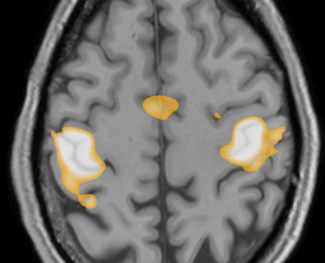
Finger movement generates a large change in BOLD signal (Blood Oxygen Level Dependent signal) in the “hand knob” on the primary motor cortex.
Tongue movement generates BOLD signal changes more ventrally on the motor homonculous, as seen below:
Speech, and in particular the word-generation task, localizes to Broca’s area. This is most commonly found on the left, but rarely (as in the unusual example below) can be bilateral with a large component on the right. The right-sided dominance of speech in this right-handed patient was confirmed with WADA testing.

