Neoplasms of the astrocytes (astrocytomas), are the most common form of primary brain neoplasm. The prognosis for astrocytomas depends on their grade and is better for low grade than high grade neoplasms.
Diagnosis: Low grade astrocytoma
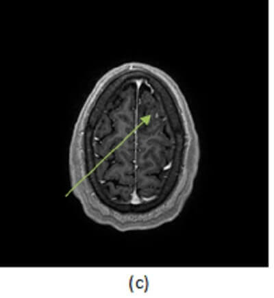
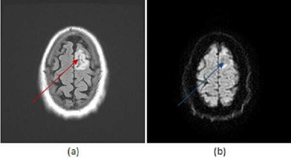
Figure 1: axial FLAIR (a, red arrow), DWI (b, blue arrow) and T1 post contrast (c ,green arrow) images of the brain reveal a circumscribed non restricting, non enhancing T2/FLAIR hyperintense mass in the high left frontal lobe.
Low grade astrocytomas are well differentiated intra-axial neoplasms. They typically present as circumscribed homogenous T2/FLAIR hyperintense supratentorial masses centered in the white matter without enhancement or restricted diffusion, although these tumors are infiltrating and microscopic disease is usually present beyond the margins apparent on imaging. There may be surrounding vasogenic edema and expansion of the cortex. Enhancement and necrosis are atypical and suggest progression to higher tumor grade such as anaplastic astrocytoma or glioblastoma multiforme.
On pathology, these tumors have moderately increased cellularity and minimal mitotic activity without microvascular proliferation or necrosis.
Treatment of low grade astroctyoma involves surgery and adjuvant radiotherapy and chemotherapy. Mean age of onset is 20-45 years. Younger age at onset, extent of resection, absence of higher grade features and supratentorial location are associated with better prognosis.