Dural arterio-venous fistulas are a rare but important category of intracranial vascular abnormalities. They are characterized by arterio-venous shunting within the dura and are most commonly a sequelae of trauma. The hypothesized pathogenesis for this diagnosis is that an inciting event like trauma leads to thrombosis of the venous sinus’, most commonly the transverse. Neoangiogenesis resulting from this trauma leads to a network of collaterals arising from the wall of the vein which ultimately anastomose with arterial collaterals on the pial (for a pial AVF) or dural (for a dural AVF) surface.
Diagnosis: Dural arterio-venous fistula (Dural AVF)
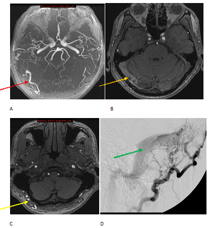
Figure 1: A-C maximum intensity projection (A) and source images (B-C) from an MR angiogram demonstrate enlargement of the right occipital artery (red arrow) with a tangle of interosseus vessels (yellow arrow) and early flow related enhancement in the transverse sinus (orange arrow) . The constellation of findings is most consistent with an arterio-venous fistula. Lateral projection (D) from a cerebral angiogram demonstrates enlargement of the occipital artery and early opacification of the transverse sinus (green arrow) consistent with an arterio-venous fistula. No tangle of vessels is seen to suggest an arterio-venous malformation.
In the evaluation of AVF’s, it is important to look for adjacent complications, especially parenchymal hemorrhage, venous infarcts and sinus thrombosis.
Morbidity of a dural AVF is primarily determined by the risk of hemorrhage and is graded using conventional angiography according to the Cognard classification. Dural AVF’s located in the sinus wall with no reflux and normal antegrade venous drainage have a low risk of hemorrhage; Those with extensive venous reflux and ectasia with involvement of the cortical veins have a hemorrhage risk near 65% and are treated, typically with embolization using a glue agent such as onyx (see figure 2). It is important to keep in mind that dural AVF’s can also occur within the spine where they present with variable but progressive myelopathy.

Figure 2: Dural AVF seen in figure 1 treated with glue embolization. Note the lack of an early draining vein.
Selected References
Imaging of dural arteriovenous fistula. Morris JM. Radiol Clin North Am. 2012 Jul; 50(4):823-39. Review.