Arteriovenous malformations (AVM’s) are vascular malformations caused by an abnormal communication (arteriovenous shunting) between small arteries and veins without an intervening capillary bed. AVM’s are the most common symptomatic intracranial vascular malformation, associated with hemorrhage in about 50% of cases. They typically present as a tangled nidus of small arteries and veins with one or more feeding arteries and draining veins. These draining veins become arterialized due to high flow leading to varices and aneurysms.
Diagnosis : Arteriovenous malformation (AVM)
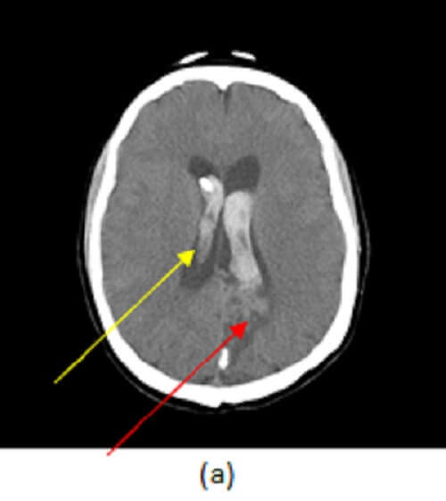
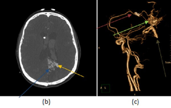
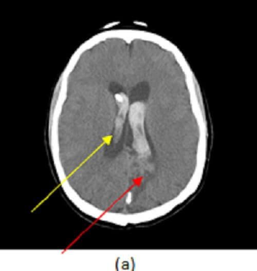
Figure 1: (a) axial image from a non contrast CT demonstrates intraparenchymal (yellow arrow) and intraventricular (red arrow) hemorrhage. (b, blue arrow), axial post contrast maximal intensity projection image from a CTA demonstrates a tangled vascular mass in the left parietal lobe consistent with an arteriovenous malformation (AVM). A saccular outpouching consistent with an intranidal aneurysm is also present adjacent to the ventricle (orange arrow). (c) Shaded surface display images show the AVM (green arrow) with early draining veins in to the deep (purple arrow) and superficial (white arrow) venous system. The intranidal aneurysm is also identified (brown arrow).
Nontraumatic hemorrhage is the most common complication of an AVM and is most commonly intraparenchymal. The risk of hemorrhage increases with age and is on the order of 2-4% per year.
AVM’s may be treated through embolization, resection or radiosurgery. The Spetzler Martin scale is commonly used to evaluate neurosurgical risk. Risk factors include AVM size greater than 3 cm, drainage of the AVM into the deep venous system (e.g. internal cerebral veins, vein of galen, straight sinus) and location in eloquent cortex (e.g. somatosensory, motor cortex, language area).