Aberrant course of the internal carotid artery (a-ICA) is a congenital anomaly characterized by the displacement of the ICA posterior and lateral to its expected course. It results from the agenesis of the C1 segment of the distal ICA during embryogenesis, leading to the absence of the vertical portion of the carotid canal and enlargement of a collateral vessel -the “aberrant” ICA- which runs posterior to anterior through middle ear across the cochlear promontory to anastomose with the petrous ICA via a dehiscent carotid plate.
Diagnosis: Left aberrant carotid artery
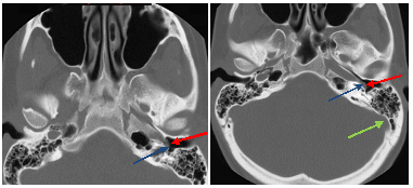
Figure 1: Axial images from a temporal bone CT demonstrate a tubular soft tissue mass (red arrow) inseparable from the petrous portion of the internal carotid, overlying the cochlear promontory (blue arrow) and directed posterior to anterior. The sigmoid sinus (green arrow) is unremarkable.
Imaging demonstrates absence of the vertical portion of the carotid canal, an intact petrous carotid and an atypical course of the ICA and a middle ear soft tissue mass. The diagnosis can be confirmed with a CT or MR angiogram which demonstrates the vascular nature of this mass.
a-ICA is a rare cause of pulsatile tinnitus, a throbbing, cyclical wooshing or buzzing sound that can be either objective (heard through auscultation) or subjective . The most common finding on physical exam is of a red coloured / vascular retrotympanic middle ear mass.
Other causes of retrotympanic masses include glomus tympanicum, middle ear schwannoma; glomus tympanicum can also appear vascular. A-ICA is an important diagnosis to exclude in this context since a biopsy can lead to uncontrollable hemorrhage. Management of a-ICA is typically expectant unless associated with an aneurysm or hemorrhage in which case coil embolization may be considered.
Selected References:
Swartz J, Loevner L. Imaging of the Temporal Bone. 4th Ed. Thieme New York 2009