In this case, images show a congenital malformation in which the corpus callosum, which connects the left and right cerebral hemispheres, failed to develop. While this abnormality is rare, there are many potential causes: in this case it was associated with a chromasomal duplication. The corpus callosum develops between the 21st and 84th day of gestation, so non-genetic insults during this period can also cause hypogenesis or complete agenesis.
Diagnosis: Corpus Callosum Agenesis
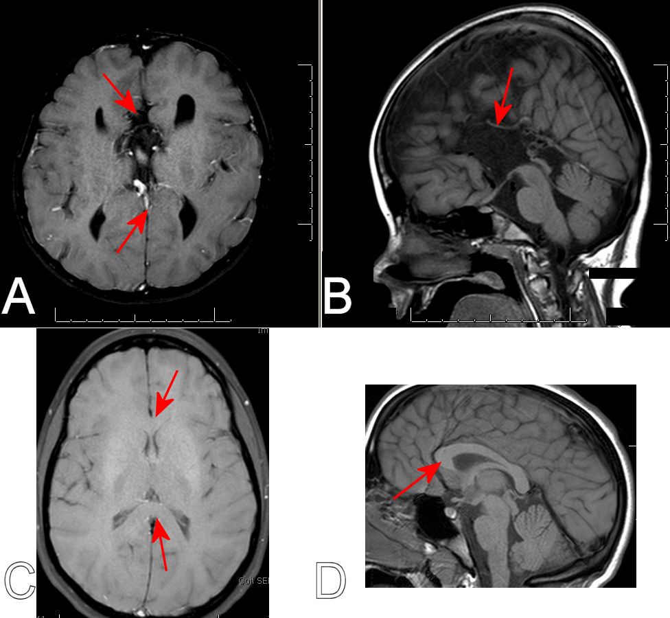
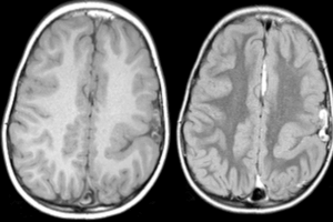
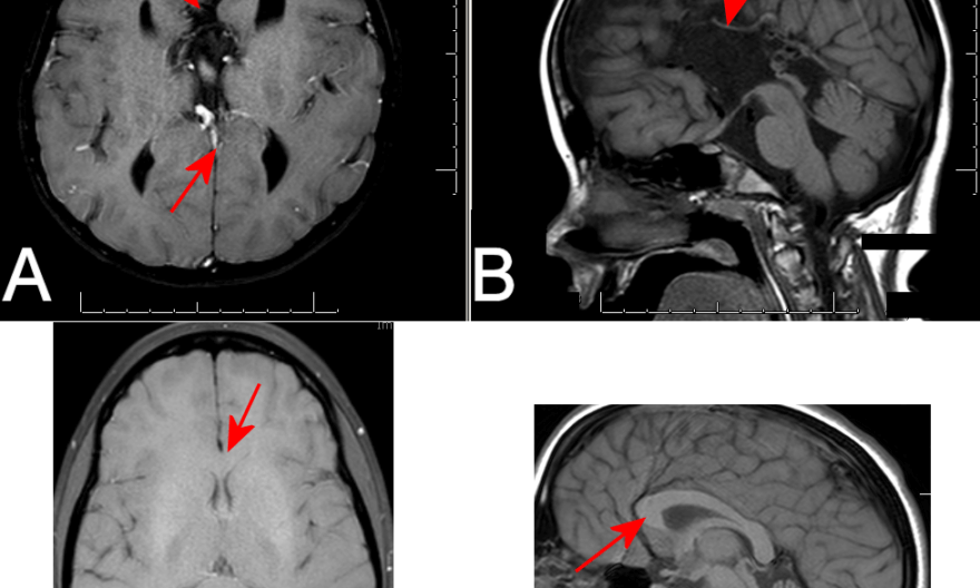
Figure 1: A: Axial T1 image showing no intra-hemispheric connections. B. A mid-sagittal cut showing no evidence of a corpus callosum. Arrows in A and B show where the corpus callosum would normally be found. C and D are shown for comparison, and demonstrate a thick corpus callosum connecting the two hemispheres.
Definitive diagnosis requires neuroimaging, but there are signs of corpus callosum agenesis on EEG. Consistently asymmetric activity in the two hemispheres is a hint, but is not specific. However, normal sleep spindles are symmetric across the hemispheres – when sleep spindles consistently occur independently in the right and left hemisphere there is almost always a missing corpus callosum.
There are some cases in which a very thin corpus callosum exists but is difficult to visualize. In these cases, the gyrus immediately adjacent to the thin corpus callosum would run rostro-caudally. In cases that truly have no corpus, like that shown above, the gyri tend to run perpendicular to where the corpus callosum would normally sit.