
Sleep stages can be identified by the appearance of characteristic electrical patterns on EEG. Sleep can initially be broken down into REM-sleep and non-REM sleep, with further parcelization of non-REM sleep into stages 1-4.
Stage 1 of non-REM sleep is the state of being drowsy, while stage 2 usually constitutes just more than 50% of nighttime sleep in the young adult. Stages 3 and 4 together form “slow-wave” sleep and are characterized by progressively more low-frequency content.
Stage 1 sleep
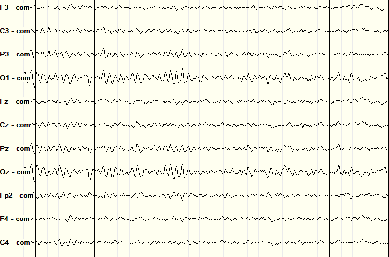
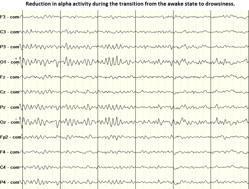
The transition from wakefulness into stage 1 sleep is heralded by the loss of alpha-range activity. The rarely happens abruptly, more often presenting as a fragmentation of the alpha rhythm with subsequent progressive decrease in epochs (Figure 1).
Figure 1: loss of alpha-frequency activity at the transition between wakefulness and stage 1 sleep.
If the EEG low-frequency filter is set very low, it might also be possible to see low frequency (0.25-0.5 Hz) variations in frontal leads that correspond to slow rolling eye movements (SREM). However, most EEG systems are set with default low-frequency filters from 0.3 to 1 Hz so it is rare see SREMs without explicitly looking for them.
Stage 1 and 2 sleep
There are two distinct patterns common to both stages 1 and 2, the are vertex waves and POSTS.
Vertex sharp-waves are found in the midline scalp electrodes, most commonly CZ. The vertex waves are about 100-200 ms in duration.
Figure 2: Vertex wave on EEG

The acronym POSTS stands for Positive Occipital Sharp Transients of Sleep. These are sharp waves found in stages 1 and 2 of sleep that typically come in runs of a one to a few seconds at a time and are found, as the name implies, in the occipital regions. POSTS are age-dependent, found infrequently in children 4-5 years of age but more frequently in teenagers and adults through age 50, after which they are less common. They have medium amplitudes (50-100 microvolts) and look like “reverse check marks.”
Figure 3: POSTS of non-REM sleep

Stage 2 sleep
The presence of sleep spindles defines stage 2 sleep. Spindles are runs of beta-frequency (~15 Hz) activity for 1-2 seconds at a time across almost all leads (see Figure 4).
K-complexes consist of large amplitude, low frequency bimodal waves typically affiliated with sleep spindles. The K-complex lasts anywhere from 1/3 to 2/3 of a full second. While K-complexes are largest along the midline, they can often be seen, to some extent, in nearly all the leads of the EEG because they are such large amplitude. An example of a K-complex and associated spindle is shown in Figure 4.
Figure 4: K-complex and sleep spindles of stage 2 sleep

Slow-wave sleep: stages 3 and 4 of non-REM sleep
Delta-wave activity (<4 Hz) defines slow wave sleep, which is composed of stages 3 and 4 on non-REM sleep. The primary factor differentiating stage 3 from stage 4 sleep is the amount of delta frequency present. In stage 3 delta activity occupies only 20-50% of the time while in stage 4 delta activity occupies 50% or more.
Slow wave sleep is important for cortical plasticity and learning (Frank et al. 2001), and the amount of slow wave sleep shrinks with age.
Figure 5: delta-frequency activity during slow-wave sleep

References
- Selim R Benbadis, MD, Diego Rielo, MD (2102) Normal Sleep EEG. Medscape.
- Frank MG, Issa NP, Stryker MP. Sleep enhances plasticity in the developing visual cortex. Neuron. 2001 Apr;30(1):275-87.