Herpes simplex virus type 1 (HSV-1) is a prevalent virus with an affinity for the nervous system. Reactivation of the virus within the brain parenchyma either directly or through retrograde spread along the cranial nerves (especially trigeminal and olfactory) can result in a severe encephalitis that is universally fatal without early treatment. In fact, HSV is the most common cause of fatal encephalitis worldwide and in the US and is associated with significant morbidity even with appropriate treatment.
Diagnosis: Herpes Encephalitis


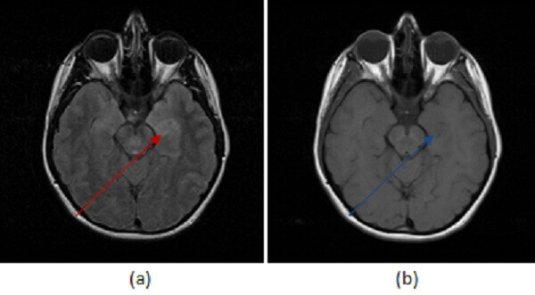
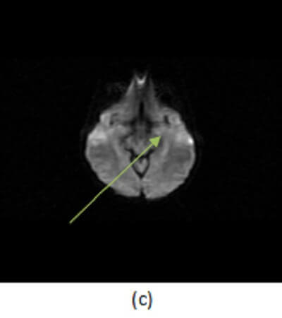
Figure 1: Increased (a,red arrow) FLAIR signal abnormality preferential for the left mesial temporal lobe is T1 hypointense (b, blue arrow ). It does not demonstrate restricted diffusion (c, green arrow) or enhance (not shown).
Clinical symptoms include a rapid onset of fever, headache,neck stiffness, photophobia, altered mental status seizures, focal neurologic signs, and impaired consciousness. Lumbar puncture classically demonstrates increased opening pressure, normal glucose, elevated protein and mild pleocytosis.
Understanding the imaging features of HSV encephalitis is important, since empiric antiviral therapy should be initiated if there is any suspicion for the condition. MRI is the most sensitive imaging modality. Imaging findings include T2/FLAIR signal abnormality involving the cortical and subcortical regions within the limbic system, especially the mesial temporal lobe. The findings are possibly bilateral but commonly asymmetric. There may be associated restricted diffusion on DWI and hemorrhage suggested by foci of decreased signal intensity on GRE sequences. Patchy or gyriform enhancement may be seen in these regions after contrast administration, or there may be no enhancement at all. The differential diagnosis also includes infarct and astrocytoma. A signal abnormality that lacks territorial restricted diffusion or that crosses vascular territories in a patient with sudden onset of symptoms and a suggestive LP help to differentiate HSV from infarct and tumor.

